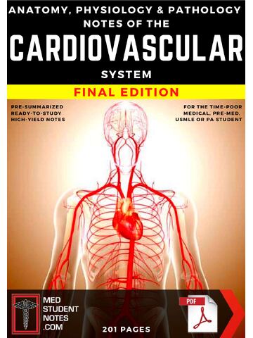
Anatomy, Physiology & Pathology Notes of the CARDIOVASCULAR SYSTEM Final Edition | PRE-SUMMARIZED READY-SUMMARIZED HIGH-YIELD NOTES For NURSING, MEDICAL, PRE-MED, USMLE OR PA STUDENT
$19.00
Secure checkout
Instant digital download
PDF document
Document details
- Pages
- 199
- File size
- 8.8 MB
- Format
- Digital PDF
- Course
- Medicine
- Category
- Study Notes
Sign in or create a free account to continue. Your purchase will be saved in My Downloads.
About this ebook
1. Anatomy of the Heart
- Layers of the Heart Wall:
- Epicardium: Outer protective layer (visceral layer of serous pericardium).
- Myocardium: Middle muscular layer; responsible for pumping. Thickest in the left ventricle.
- Endocardium: Inner endothelial lining of heart chambers and valves.
- Chambers & Blood Flow:
- Right Atrium (RA): Receives deoxygenated blood via Superior/Inferior Vena Cava and Coronary Sinus.
- Right Ventricle (RV): Pumps deoxygenated blood through the pulmonary valve into the pulmonary trunk/arteries.
- Left Atrium (LA): Receives oxygenated blood via four pulmonary veins.
- Left Ventricle (LV): Pumps oxygenated blood through the aortic valve into the aorta. (3x thicker muscle wall than RV).
- Valves:
- Atrioventricular (AV) Valves: Tricuspid (right) and Bicuspid/Mitral (left). Prevent backflow into atria during ventricular systole. Anchored by chordae tendineae and papillary muscles.
- Semilunar (SL) Valves: Aortic and Pulmonary. Prevent backflow into ventricles during ventricular diastole.
2. Cardiac Physiology & Electrical Conduction
- Conduction System Pathway:
- SA Node (Sinoatrial): Primary pacemaker (60–100 bpm). Located in the upper right atrium.
- AV Node (Atrioventricular): Delays impulse (~0.1 sec) to allow atrial contraction/filling (40–60 bpm).
- Bundle of His (Atrioventricular Bundle): Transmits signal down interventricular septum.
- Right & Left Bundle Branches: Conduct impulses toward apex.
- Purkinje Fibers: Rapidly spread impulse through ventricular myocardium (20–40 bpm).
- The Cardiac Cycle (Wiggers Diagram Basics):
- Systole: Contraction phase (isovolumetric contraction + ventricular ejection).
- Diastole: Relaxation phase (isovolumetric relaxation + ventricular filling).
- S1 ("Lub"): Closure of AV valves (mitral/tricuspid) at the start of systole.
- S2 ("Dub"): Closure of SL valves (aortic/pulmonic) at the end of systole.
- Hemodynamic Formulas:
- Cardiac Output (CO): CO = HR × SV (Normal: ~5 L/min).
- Stroke Volume (SV): End-Diastolic Volume (EDV) minus End-Systolic Volume (ESV). Determined by:
- Preload: Degree of myocardial stretch before contraction (venous return).
- Afterload: Resistance the ventricle must overcome to eject blood (aortic pressure/SVR).
- Contractility: Inotropy; intrinsic strength of cardiac muscle.
- Mean Arterial Pressure (MAP): \(\text{MAP} = \text{DBP} + \frac{1}{3}(\text{SBP} - \text{DBP})\) or CO × SVR.
3. Vascular Physiology & Blood Pressure
- Vessel Types:
- Elastic Arteries (Aorta): Windkessel effect; smooth out pressure surges.
- Muscular/Distribution Arteries: Adjust blood flow via vasoconstriction/vasodilation.
- Arterioles: Resistance vessels; primary controllers of systemic blood pressure.
- Capillaries: Exchange sites (single layer of endothelial cells).
- Veins/Venules: Capacitance vessels; hold ~64% of blood volume.
- Regulation of Blood Pressure:
- Baroreceptors: Carotid sinus and aortic arch detect stretch; send signals to medulla to adjust HR and vasomotor tone.
- RAAS (Renin-Angiotensin-Aldosterone System): Low BP → Kidney releases renin → Angiotensinogen converted to Angiotensin I → ACE converts to Angiotensin II (potent vasoconstrictor) → Aldosterone release → Na⁺ and H₂O reabsorption.
4. Pathology & High-Yield Disease States
- Ischemic Heart Disease (IHD):
- Angina Pectoris: Transient chest pain without necrosis. Stable (predictable) vs. Unstable (unpredictable/crescendo).
- Myocardial Infarction (MI): Necrosis of myocardial tissue due to prolonged ischemia.
- STEMI (ST-elevation) vs. NSTEMI (non-ST elevation).
- Biomarkers: Troponin I/T (rises in 2–4 hrs, peaks at 24 hrs, stays elevated 7–10 days) and CK-MB.
- Heart Failure (HF):
- Left-Sided HF: Backup into lungs. Symptoms: Dyspnea on exertion, orthopnea, paroxysmal nocturnal dyspnea, crackles, S3 gallop.
- Right-Sided HF: Backup into systemic circulation. Symptoms: Jugular venous distension (JVD), peripheral pitting edema, hepatomegaly, ascites. (Most common cause is left-sided HF).
- Valvular Disorders:
- Aortic Stenosis: Obstruction of LV outflow. Causes systolic ejection murmur (crescendo-decrescendo). Classic triad: Syncope, Angina, Dyspnea (SAD).
- Mitral Regurgitation: Backflow into LA during systole. Causes holosystolic/pansystolic murmur radiating to the axilla.
- Hypertension (HTN) & Vascular Pathology:
- Essential vs. Secondary HTN: Essential is idiopathic (90%); secondary has an identifiable cause (e.g., renal artery stenosis).
- Atherosclerosis: Accumulation of lipid-rich plaques in the intima of large/medium arteries → risk of rupture, thrombosis, aneurysm.
File included
Anatomy-Physiology-Pathology-Notes-of-the-CARDIOVASCULAR-SYSTEM-Final-Edition.pdf
8.8 MB
Related ebooks