
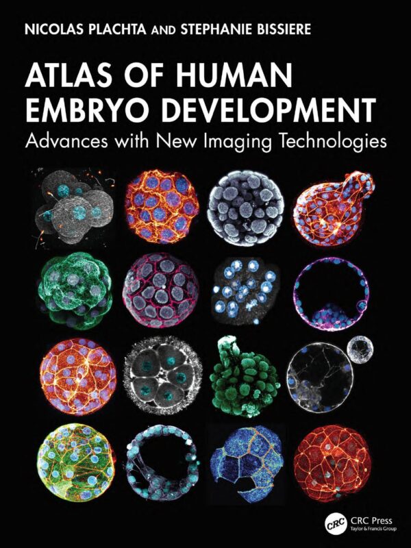
Atlas of Human Embryo Development Advances with New Imaging Technologies 1st Edition By Nicolas Plachta, Stephanie Bissiere
$30.00
Secure checkout
Instant digital download
PDF document
Document details
- Pages
- 121
- File size
- 5.16 MB
- Format
- Digital PDF
- Course
- Medicine
- Category
- eBook[PDF]
Sign in or create a free account to continue. Your purchase will be saved in My Downloads.
About this ebook
The Atlas of Human Embryo Development: Advances with New Imaging Technologies (1st Edition), written by Nicolas Plachta and Stephanie Bissiere (published by CRC Press / Routledge), focuses strictly on the early visual stages of human life. [1]
The primary topics covered in this atlas include:
- Fertilization to Pre-Implantation: A comprehensive timeline showing the biological progression from the moment of fertilization to the final stages of pre-implantation blastocyst development. [1, 2]
- Subcellular Embryogenesis: Visualizing dynamic intracellular and structural changes that drive early embryogenesis, captured in 3D and in real time. [1, 2]
- Developmental Defects: Identification of early visual anomalies, impaired development, and mitotic or segregation errors that lead to congenital and genetic conditions. [1, 2, 3]
- Advanced Medical Imaging Techniques: Documentation of non-invasive live-imaging technologies (such as membrane-permeable fluorescent dyes) that replace traditional fixed-specimen light microscopy. [1]
- Clinical Applications in Reproductive Medicine: Practical insights aimed at improving IVF success rates, monitoring embryo quality, and advancing fertility treatments. [1]
File included
tmpphpS3Crnv
5.16 MB
Related ebooks