
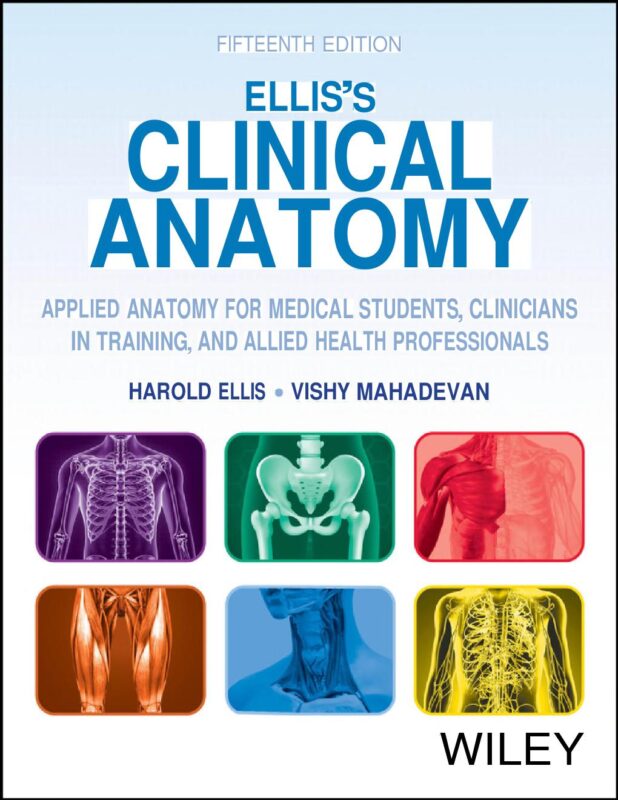
Elliss Clinical Anatomy Applied Anatomy for Medical Students Clinicians in Training and Allied Health Professionals 15th Edition By Harold Ellis, Vishy Mahadevan
$25.00
Secure checkout
Instant digital download
PDF document
Document details
- Pages
- 842
- File size
- 31.79 MB
- Format
- Digital PDF
- Course
- Education
- Category
- eBook[PDF]
Sign in or create a free account to continue. Your purchase will be saved in My Downloads.
About this ebook
The Ellis’s Clinical Anatomy: Applied Anatomy for Medical Students, Clinicians in Training, and Allied Health Professionals by Harold Ellis and Vishy Mahadevan is systematically organized into six core regional sections. [1]
The book focuses heavily on functional anatomy, surface markings, and the precise anatomical basis required for practical medical procedures and surgical interventions. [1]
The core regional topics covered across its sections include: [1]
Part 1: The Thorax
- The Thoracic Wall: Ribs, intercostal spaces, internal thoracic vessels, and breasts.
- The Pericardium and Heart: Chambers, valves, blood supply, nerve supply, and developmental anomalies.
- The Mediastinum: Structures of the superior, anterior, middle, and posterior mediastinum including the trachea, esophagus, and thoracic duct.
- The Lungs and Pleurae: Pleural reflections, bronchopulmonary segments, and the mechanics of respiration.
Part 2: The Abdomen and Pelvis
- The Anterior Abdominal Wall: Muscles, fascial layers, inguinal canal, and abdominal hernias.
- The Peritoneum: Peritoneal cavity, lesser sac, and mesenteries.
- The Gastrointestinal Tract: Stomach, small intestine, large intestine, appendix, and rectum.
- The Visceral Organs: Liver, gallbladder, pancreas, and spleen.
- The Urinary Tract: Kidneys, ureters, bladder, and urethra.
- The Reproductive System: Male and female internal and external genitalia, pelvic floor, and perineum.
Part 3: The Upper Limb
- The Shoulder Girdle: Bones, joints, axilla, and the brachial plexus.
- The Arm and Forearm: Compartments, muscle groups, cubital fossa, and neurovascular structures.
- The Hand: Fascial spaces, tendons, intrinsic muscles, and functional mechanics of the fingers.
Part 4: The Lower Limb
- The Hip and Thigh: Gluteal region, femoral triangle, hip joint, and main nerve paths (femoral, sciatic, obturator).
- The Knee and Leg: Knee joint stabilizers, popliteal fossa, and compartments of the leg.
- The Foot and Ankle: Ankle joint, arches of the foot, tarsal tunnel, and retinacula.
Part 5: The Head and Neck
- The Neck: Fascial layers, triangles of the neck, thyroid gland, parathyroid glands, and main vessels.
- The Face and Scalp: Muscles of facial expression, mastication, parotid gland, and blood supply.
- The Cranial Cavity: Meninges, dural venous sinuses, and intracranial pathways.
- Special Senses & Airway: Orbit and eye, ear, nasal cavity, paranasal sinuses, pharynx, and larynx.
Part 6: The Nervous System
- The Brain: Cerebral hemispheres, basal ganglia, thalamus, brainstem, and cerebellum.
- The Spinal Cord: Tracts, blood supply, and lumbar puncture anatomy.
- The Cranial Nerves: Complete clinical anatomy and pathway courses for CN I through CN XII.
- The Autonomic Nervous System: Sympathetic and parasympathetic pathways.
File included
tmpphpl35P3j
31.79 MB
Related ebooks