
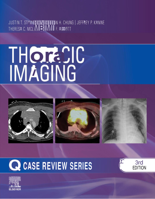
Thoracic Imaging: Case Review 3rd Edition By Justin Stowell, Jonathan Chung, Jeffrey Kanne, Theresa McLoud, Gerald Abbott
$30.00
Secure checkout
Instant digital download
PDF document
Document details
- Pages
- 250
- File size
- 21.41 MB
- Format
- Digital PDF
- Course
- Education
- Category
- eBook[PDF]
Sign in or create a free account to continue. Your purchase will be saved in My Downloads.
About this ebook
Thoracic Imaging: Case Review, 3rd Edition, authored by Justin T. Stowell, Jonathan H. Chung, Jeffrey P. Kanne, Theresa C. McLoud, and Gerald F. Abbott, is a case-based study guide published by Elsevier in July 2023. It is designed for radiology residents and recertifying radiologists preparing for board exams. [1, 2, 3]
Key Features
- Over 145 to 200 high-yield case studies.
- Organized by level of difficulty (mild, moderate, hard).
- Multiple-choice questions, answers, and rationales styled after certification tests.
- High-quality chest X-rays, CT scans, PET-CT, and MRIs. [1, 2]
Covered Topics
- Lung cancer and pulmonary nodules (including Fleischner guidelines).
- Idiopathic interstitial pneumonias and small airway disease.
- Sarcoidosis, lymphoma, and traumatic aortic injury.
- Emerging pulmonary infections and smoking-related lung disease. [1, 2]
The book contains roughly 150 to 200 high-yield clinical cases split across three core operational sections: [1, 2, 3]
1. Opening Round (Easy Cases)
This introductory section establishes baseline core knowledge. It focuses on primary diagnostic interpretations, expected lines, and routine critical care findings. [1, 2]
- Basic Trauma: Rib fractures, pneumothorax, and minor hemothorax.
- Lines and Tubes: Evaluation of endotracheal tube placement, central venous catheters, and enteric tubes.
- Common Neoplasms: Typical presentations of clear-cut lung masses and benign nodules.
- Classic Infections: Standard community-acquired bacterial pneumonias. [1, 2]
2. Fair Game (Moderate Cases)
The intermediate section tests your proficiency with everyday clinical scenarios that require differential diagnosis. [1]
- Diffuse Lung Disease: Initial assessment of interstitial patterns using HRCT.
- Vascular Anomalies: Uncomplicated pulmonary embolisms and stable aortic ectasia.
- Pleural and Mediastinal Diseases: Standard pleural effusions, loculated collections, and common anterior mediastinal masses (e.g., thymoma).
- Nodule Management: Applying the Fleischner guidelines to evaluate solitary pulmonary nodules. [1, 2, 3]
3. Challenge Round (Hard Cases)
The advanced section presents complex multi-system disorders and atypical disease patterns requiring sophisticated diagnostic pearls. [1, 2]
- Advanced Vascular Pathology: Traumatic aortic injuries, complex dissection patterns, and severe pulmonary hypertension.
- Atypical & Emerging Infections: Fungal, viral, or opportunistic infections in immunocompromised hosts.
- Complex Interstitial Lung Diseases (ILDs): Idiopathic interstitial pneumonias, sarcoidosis, and occupational pneumoconioses.
- Smoking-Related and Small Airway Conditions: Confounding presentations of smoking-related lung injuries. [1, 2, 3]
File included
eBook-Thoracic-Imaging-Case-Review-3e-Justin-Stowell.pdf
21.41 MB
Related ebooks