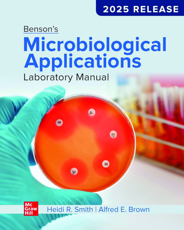
Benson’s Microbiological Applications Laboratory Manual 2025 Release 15th Edition By Heidi Smith, Alfred Brown
$30.00
Secure checkout
Instant digital download
PDF document
Document details
- Pages
- 481
- File size
- 31.87 MB
- Format
- Digital PDF
- Course
- Education
- Category
- eBook[PDF]
Sign in or create a free account to continue. Your purchase will be saved in My Downloads.
About this ebook
The Benson's Microbiological Applications Laboratory Manual (2025 Release / 15th Edition), authored by Heidi Smith and Alfred E. Brown, covers all foundational and advanced topic areas required for general and undergraduate microbiology laboratory courses. Published by McGraw Hill, this 480-page manual is structured into targeted sections containing step-by-step experimental exercises. [1, 2, 3, 4]
Core Laboratory Sections & Topics Covered
🔬 Part 1: Microscopy
- Brightfield Microscopy: Principles of magnification, resolution, and standard operation.
- Darkfield Microscopy: Visualising live, unstained specimens against a dark background.
- Phase-Contrast Microscopy: Enhancing contrast in transparent and internal cellular structures.
- Microscopic Measurements: Calibrating and utilizing ocular and stage micrometers. [1]
🦠 Part 2: Survey of Microorganisms
- Microbiology of Pond Water: Identification of protists, microalgae, and cyanobacteria.
- Ubiquity of Bacteria: Demonstrating environmental distribution and surface contamination.
- The Fungi: Morphological classification of molds and yeasts. [1]
🧫 Part 3: Manipulation of Microorganisms (Aseptic Technique)
- Aseptic Transfer: Techniques for moving cultures without introducing contamination.
- Streak Plate Method: Isolation of pure colonies from mixed cultures.
- Pour Plate Method: Quantifying and isolating microbes via serial dilutions. [1, 2]
🎨 Part 4: Staining and Observation of Microorganisms
- Smear Preparation: Fixing bacterial slides safely.
- Simple Staining: Visualising cellular morphology and arrangement using a single dye.
- Negative Staining: Observing fragile structures without heat-fixing.
- Gram Staining: Differentiating between Gram-positive and Gram-negative cell walls.
- Endospore & Acid-Fast Staining: Diagnostic staining for highly resistant structures. [1, 2]
🧪 Part 5: Culture Methods & Environmental Influences
- Culture Media: Formulating simple, complex, selective, and differential media.
- Physical Factors: Effects of temperature, pH, osmotic pressure, and oxygen requirements on growth.
- Chemical Control: Testing the efficacy of antiseptics, disinfectants, and antibiotics via disk diffusion. [1, 2]
🧬 Advanced Sections (Complete Version Modules)
- Bacterial Viruses: Isolation and titration of bacteriophages.
- Microbial Genetics: Bacterial transformation, conjugation, and basic DNA manipulation.
- Medical Microbiology: Identification of unknown pathogens using selective tests, biochemical profiles, and rapid diagnostic kits. [1, 2]
File included
eBook-Bensons-Microbiological-Applications-Laboratory-Manual-2025-Release-By-Heidi-Smith-Alfred-Brown.pdf
31.87 MB
Related ebooks